Overview
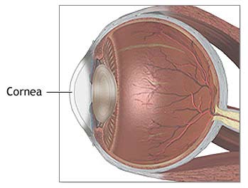
The cornea is the clear, dome-shaped tissue on the front of the eye that covers the pupil and iris. It is an integral part of the visual system as it provides the majority of the focusing power of the eye onto the retina to form a clear and crisp image. Maintaining a healthy cornea is therefore a very important part of maintaining good overall eye health and vision.
There are many diseases that can affect the cornea, including infections, genetic conditions, dry eye disease, and traumatic injuries. Here at Caplan Eye Clinic, we are equipped with the latest in technological instruments, medical treatments, and surgical techniques to care for corneal disease.

Dry Eye Syndrome
Also known as Tear Film Dysfunction, dry eye syndrome is one of the most common eye conditions affecting the general population. Symptoms can include blurred or fluctuating vision, redness, foreign body sensation, discharge, eye pain, and tearing. As its name implies, dry eye syndrome is caused by an unhealthy or unstable tear film on the surface of the cornea. There are many causes of dry eye, including normal aging, environmental factors, systemic medications, medicated eye drops, eyelid inflammation, contact lens use, and certain medical problems.
Dry eye syndrome can be a difficult problem to treat and often becomes a chronic issue. Treatment should be aimed at reducing the severity of the symptoms and preventing the more serious complications that can arise in advanced cases. Numerous over-the-counter treatments are available to relieve the milder symptoms of dry eye. More advanced treatments can be performed in the office, including plugging the drainage ducts for the tears in the eyelids. There are also prescription-strength eye drops that reduce inflammation on the surface of the eye and promote the production of healthy tears.
Keratoconus
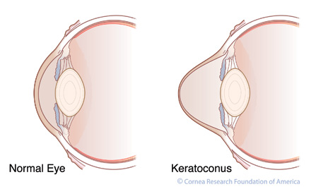
 Keratoconus is a progressive disease of the cornea in which the central part of the cornea becomes thinner and starts to bulge. This bulging causes the cornea to take on a cone-like shape instead of the usual dome-shape. The misshapen cornea cannot focus light properly, leading to blurred vision. Keratoconus usually affects patients in their teens and twenties but can occur at any age. Besides blurred vision, symptoms can also include glare, light sensitivity, and inability to tolerate contact lenses. The causes of keratoconus are unknown, but chronic eye rubbing can contribute to the disease.
Treatment early in the disease process can include eyeglasses or hard contact lenses to correct the astigmatism caused by keratoconus. An exciting new treatment currently undergoing FDA trials in the United States is collagen crosslinking. Although not a cure for keratoconus, this procedure can halt the progression of the disease and has been performed for many years overseas in Europe and South America. For severe cases of keratoconus, corneal transplantation may be required, although this is uncommon. Image: Keratoconus.jpeg (www.cornea.org)
Fuchs’ Corneal Dystrophy
Fuchs’ Dystrophy is a genetic condition affecting the inner layer of the cornea called the endothelium. This layer is responsible for keeping the cornea dehydrated and compact which results in its ability to stay clear. In Fuchs’ Dystrophy, the endothelium prematurely degenerates, resulting in progressive swelling of the cornea. When the cornea is swollen, it becomes cloudy and cannot focus light properly, leading to poor vision.
Initially, patients may be asymptomatic when the dystrophy is diagnosed. Early symptoms include glare, halos around lights, and blurry vision that is often worse earlier in the day. Advanced symptoms include persistently blurred and deteriorating vision.
Treatment for early Fuchs’ dystrophy includes environmental changes and eye drops to help reduce the swelling in the cornea. Advanced cases may require surgery with corneal transplantation to replace the diseased layer of the cornea with a healthy one. Historically these surgeries have required full-thickness transplants called PKPs (penetrating keratoplasty), but newer techniques have developed which only require replacement of the diseased layer of the cornea. This surgery, called a DSAEK (Descemet’s Stripping Automated Endothelial Keratoplasty), uses a partial thickness transplants and has the advantages of faster visual recovery, fewer stitches, and lower risk of graft rejection than a traditional cornea transplant.
Pterygium
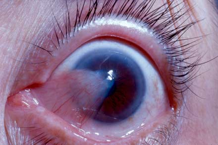
 A pterygium is a benign, wing-like growth of the conjunctiva (the clear tissue over the white wall of the eye) over the cornea, usually from the nose-side of the eye. It is commonly seen in patients who have spent a significant amount of time outdoors or grew up in warmer climates and originates from exposure to ultra-violet light, wind, and sun.
Although some may be able to see the pterygium on the surface of their eye, most patients are asymptomatic, especially when the growth is early in development. However, some may experience redness, irritation, or foreign-body sensation. As the pterygium grows, it can change the shape of the cornea, affecting vision.
Most pterygia are treated conservatively with lubrication of the surface of the eye to reduce irritating symptoms. If they become large or bothersome enough, they can be surgically removed in the operating room. Image: Pterygium.jpeg (www.surfereye.com)
Dr. Brendon Sumich is a comprehensive ophthalmologist with subspecialty training in cornea and external eye disease at the New England Eye Center and Ophthalmic Consultants of Boston in Boston, MA. He is proud to offer the latest in treatments and procedures to manage all of your corneal problems.


